

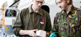


With the hope of guiding students to develop creativity and problem-solving skills, the team at the Health Design Lab surveyed their options for 3D printers and selected Ultimaker. 3D printing with Ultimakers opened the doors for students to work directly with clinicians, providing a platform for innovation-driven education. At the Health Design Lab, students have the unique opportunity to participate in current patient cases by working with radiologists to produce accurate anatomical models.
Enhancing patient care with 3D printing at Jefferson Health
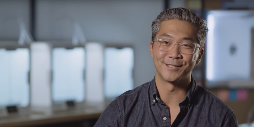
Thomas Jefferson University and Jefferson Health, a university and health system located in Philadelphia, is at the forefront of healthcare technology. When Dr. Robert Pugliese and Dr. Bon Ku set out to integrate 3D printing into the Jefferson Health Design Lab, they wanted to better prepare their students for the challenges they would face when entering real-world hospital environments.
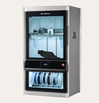
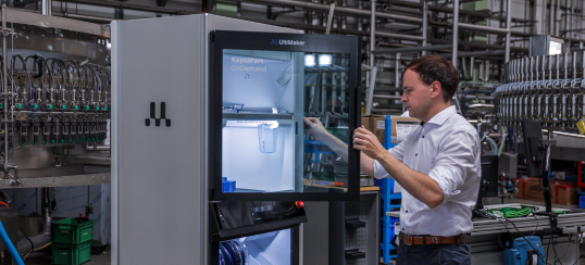
The Health Design Lab delves into a wide range of medical and educational applications, from ENT surgery to high-risk obstetrics, cardiology, ultrasound training, orthopedic surgery, and radiology. Though they first used the Ultimaker 2+ Extended, the team soon recognized the value of having multiple printheads and taking advantage of PVA water-soluble filament. After adding the Ultimaker 3, they purchased Ultimaker S5 units when the new model launched. The space is home to 14 Ultimaker 3D printers, with the Ultimaker S5 boosting speed and effectiveness for their team when they create surgical models to improve patient care.
While it is a bonus that these 3D printed patient anatomy models help the students to learn the processes behind interpreting medical scan data, segmenting critical features, and mastering the process of producing 3D printings, their efforts are also a huge benefit to the clinical practices they support. The work of the Health Design Lab to optimize their process means a fast turnaround for surgeons who are preparing for operations. “We can take an image from a patient scan, we can convert that to a 3D printed model in a matter of minutes, and then have that model in the hands of a surgeon in just about a day,” says Dr. Pugliese.
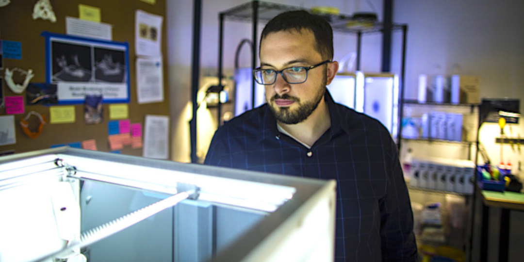
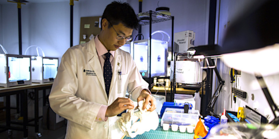
Dr. Pugliese working with the Ultimaker S5 in the Heath Design Lab
With access to the tangible models, Jefferson surgeons are able to better visualize the challenges ahead of them, providing greater insight for pre-operative planning. This is especially useful ahead of complex and high-risk procedures, providing a clearer understanding of the spatial placement of tissue and bone that can be difficult to interpret on a 2D scan.
Planning complex surgeries with 3D printed models
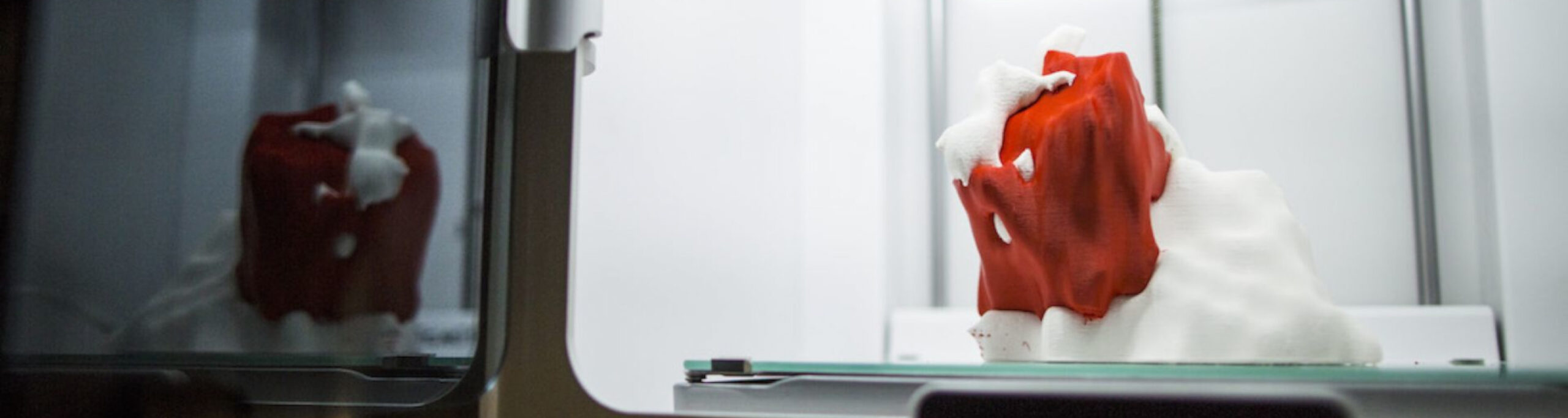
In one such case at Jefferson's Abington Hospital, a 3D printed model was used during a complicated C-section to precisely study the vasculature for a patient’s uterus. Surgeons referenced the model to identify fibroids that they would need to avoid before they made their incisions, ensuring that they minimized blood loss and reduced the risk of patient harm.
Dr. Amy Mackey, Vice Chair of the Department of Obstetrics and Gynecology at Abington Hospital, understands that these models 3D printed on Ultimaker are especially useful for patient education. “When we introduce these models to the patients their eyes get big and they ask a lot of questions, it helps them to understand what the complexity of their case really is,” she says. “It's just so much better to have the patient on the same page and these models really help bring that reality to them."
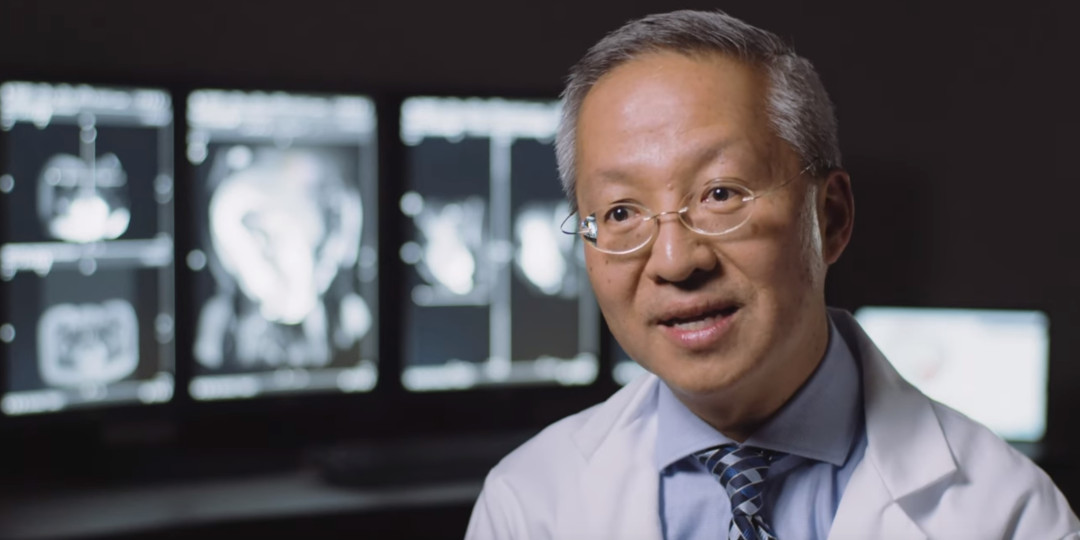
The radiology department at Abington Hospital also finds the 3D printed models useful, particularly when working with students and residents. “As a radiologist who is exposed to a lot of new technology, I'm really thrilled about showing 3D prints to my colleagues as well as to medical students and residents, because a lot of them don't know about this incredible tool that we actually have access to,” says Dr. Philip Lim, Chair of the Department of Radiology at Abington Hospital.
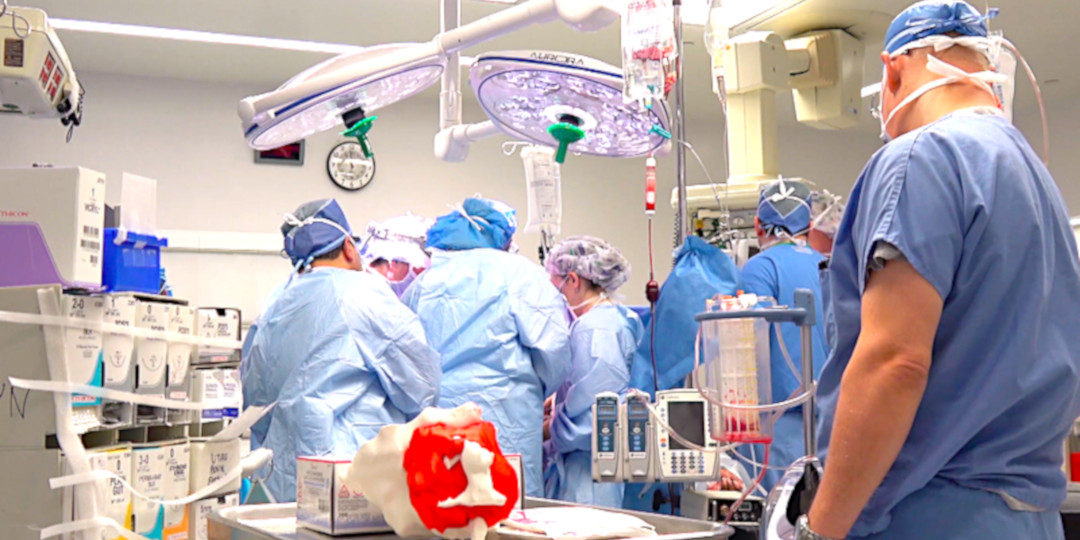
3D printed model in the operating room at Abington Hospital
According to Dr. Lim, handling the 3D printed models provides students with a greater understanding of anatomy and how they might approach a surgery themselves. It yields comfort for patients and instills confidence in their surgeons as well, demonstrating that their doctors are taking an extra step to better understand their complex case ahead of a procedure.
Tackling real-world challenges with the Ultimaker S5
Providing a valuable service to the 16 hospitals within the Jefferson Health system, Ultimaker 3D printers offer design flexibility and fast turnaround when it matters most for patient care. With four Ultimaker S5 3D printers in the Health Design Lab, the team saves time with fewer assembly and post-processing needs, because they can print large, complete anatomical models with ease.
Student reviewing a 3D printed model in the Health Design Lab
As 3D printing becomes more popular in hospitals, it will continue to have an influence on the future clinical careers of students, residents, and doctors. Impacting an array of medical applications at Jefferson Health, Ultimaker remains a natural fit for the Health Design Lab. As Dr. Ku explains, “3D printing is easy, cost-effective, and inspires my team to try new things. We chose the Ultimaker S5 because it was so reliable and easy to use, and we hope to add even more of them to our lab in the future.”
Disclaimer: Some of the statements in Ultimaker marketing materials relating to medical applications are "forward-looking" and are made pursuant to the safe harbor provision of the Private Securities Litigation Reform Act of 1995 and/or MDD 93/42 EC. These forward-looking statements include statements relating to, among other things, our planned commercialization efforts and regulatory approvals of our technologies, research and development projects as well as the success thereof, which may not be obtained in time or at all. Such statements are based on the current expectations of Ultimaker management, of which many are beyond Ultimaker management’s control and certain assumptions in light of information currently available to it. We caution you that forward-looking statements are not guarantees of future performance and involve known and unknown risks, uncertainties, and other factors that may cause our actual results, performance and/or achievements to be materially different from our expectations. Ultimaker neither intends nor assumes any obligation to update or revise these forward-looking statements in light of developments which differ from those anticipated.
Materialise Mimics inPrint is intended for use as a software interface and image segmentation system for the transfer of DICOM imaging information from a medical scanner to an output file. It is also used as pre-operative software for treatment planning. For this purpose, the Mimics inPrint output file can be used for the fabrication of physical replicas of the output file using traditional or additive manufacturing methods. The physical replica can be used for diagnostic purposes in the field of orthopedic, maxillofacial and cardiovascular applications. Mimics inPrint should be used in conjunction with other diagnostic tools and expert clinical judgment.
Please note that the 3D printed anatomical models are an assistive tool and should always be used as a complementary source to traditional diagnostic methods.
The evaluation has been performed using Ultimaker Cura 3.4, without the Toolbox. Only this version must be used printing models for diagnostic scenarios as mentioned in this printer performance validation. Only after a written explicit declamation that a newer version is validated such version may be used for the aforementioned usage.